


Everything You Need to Know About OPG X-Rays for Your Dental Health
An OPG (Orthopantomogram) X-ray is a single panoramic image that captures your entire mouth – all teeth, both jaws, and the surrounding bone structure – in one convenient scan. If your dentist has recommended an OPG or you're curious about what this imaging involves, you've come to the right place. This comprehensive guide explains what an OPG is, why dentists recommend it, what to expect during the procedure, and how to find quality X-Ray OPG Services in Bengaluru that deliver accurate results efficiently.
OPG X-rays have become the standard for comprehensive dental assessment, orthodontic treatment planning, implant evaluation, and routine dental check-ups. Unlike individual tooth X-rays that show only a small area, an OPG provides a bird's-eye view of your entire dental structure in seconds. This makes it invaluable for dentists, orthodontists, and oral surgeons who need to assess overall dental health, plan treatments, or diagnose complex issues.
What Exactly is an OPG X-Ray?
An OPG is a panoramic radiographic technique that creates a single, flat image showing all your teeth from the upper molars on one side, across the front, to the upper molars on the other side – and the same for the lower jaw. It's called "orthopantomographic" because it produces an orthoradial (undistorted) panoramic image of your dentition and supporting structures.
The imaging process is remarkably simple:
The machine rotates around your head while an X-ray source and sensor move in synchronization. As they travel, they capture a curved layer that corresponds to the dental arch, creating a comprehensive two-dimensional representation of your entire mouth in about 20 seconds.
Key characteristics of OPG imaging:
-
Single image, full mouth view – No need for multiple individual X-rays; one scan shows everything.
-
Fast and non-invasive – The procedure takes less than a minute and causes no discomfort.
-
Lower radiation dose – OPG uses less radiation than traditional full-mouth X-ray series (often 12-14 individual films).
-
Wide field of view – Shows teeth, bone levels, jaw joint (temporomandibular joint), and sinus areas.
-
Excellent for planning – Ideal for implant placement, orthodontic treatment, wisdom tooth extraction, and surgical procedures.
-
Easy patient comparison – Digital OPG images can be stored, compared over time, and shared instantly with specialists.
Why Your Dentist Recommends OPG X-Rays
Dentists recommend OPG imaging for specific clinical situations. Understanding these reasons helps you appreciate the value of the procedure and prepare appropriately:
Orthodontic Treatment Planning
Before starting braces or aligners, orthodontists use OPG X-rays to assess tooth roots, bone density, jaw growth patterns, and the position of wisdom teeth. This information is crucial for designing an effective treatment plan and predicting outcomes.
Implant Assessment
If you're considering dental implants to replace missing teeth, an OPG shows bone height and density in the proposed implant sites. This helps your implant surgeon determine feasibility and plan the procedure accurately.
Wisdom Tooth Evaluation
OPG X-rays reveal whether wisdom teeth are impacted, angled abnormally, or likely to cause problems. Oral surgeons rely on these images to plan extractions and predict complications.
Periodontal Disease Detection
Dentists use OPG images to assess bone loss around teeth, which is a hallmark of gum disease. Early detection allows intervention before severe damage occurs.
Routine Dental Check-Ups
Many dental practices obtain baseline OPG X-rays during initial consultations or periodic comprehensive exams to detect cavities between teeth, bone loss, cysts, or other pathology not visible clinically.
Trauma or Injury Assessment
After dental injuries, facial trauma, or jaw fractures, OPG X-rays help dentists and doctors assess damage and plan treatment.
Sleep Apnea and TMJ Evaluation
OPG images show the position of the lower jaw and can reveal anatomical factors contributing to sleep apnea or temporomandibular joint disorders.
Types of Dental Imaging: OPG vs. Other X-Rays
Understanding how OPG compares to other dental X-ray methods helps clarify why your dentist chose this particular imaging:
OPG (Panoramic) X-Ray
-
Shows entire mouth in one image
-
Quick, non-invasive, lower radiation
-
Best for overall assessment, planning, and screening
-
May miss fine details in specific teeth
Intraoral Periapical (PA) X-Rays
-
Shows individual teeth and roots in detail
-
High resolution, excellent for cavity detection
-
Requires multiple images for full-mouth assessment
-
Higher cumulative radiation if using many films
Bitewings
-
Captures upper and lower posterior teeth in one image
-
Excellent for detecting cavities between teeth
-
Cannot show root structure or bone levels
-
Useful for routine cavity screening
Cone Beam Computed Tomography (CBCT)
-
3D imaging showing bone structure in three dimensions
-
Highest resolution, ideal for implant planning and complex cases
-
Higher radiation dose than OPG
-
More expensive; used when OPG is insufficient
For most routine situations, OPG X-rays provide the best balance of comprehensive information, safety, and cost-effectiveness.
What to Expect During Your OPG Appointment
Knowing what happens during your OPG scan reduces anxiety and helps you prepare mentally. Here's the typical process:
Before the Scan
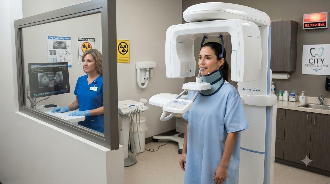
Your technician will ask you to remove jewelry, piercings, and any metal items from your head and neck area. If you have a removable denture or partial, ask whether it should be removed – policies vary. You'll be guided into the OPG machine room and positioned with your chin on a chin rest and your forehead against a head support.
Positioning is Critical
Proper positioning ensures the clearest images. Your technician will adjust the machine so the focal trough (the line of sharpness) aligns with your dental arch. You'll be asked to bite gently on a bite block and hold perfectly still. This positioning takes just a minute or two.
The Scan
Once positioned, the machine rotates around your head while you remain motionless. The entire rotation takes about 20 seconds. You'll hear quiet mechanical sounds as the arm moves. There's no pain, no sensation, and no need to hold your breath. Most people find it anxiety-free.
After the Scan
Once the image is captured, you're done. There's no recovery time. You can return to your regular activities immediately. Digital OPG images appear on the technician's screen within seconds, allowing instant quality checks before you leave.
Preparing for Your OPG X-Ray
While OPG preparation is minimal, a few simple steps ensure the best results:
-
Remove all metal jewelry – Earrings, necklaces, nose rings, tongue piercings, and hairpins must be removed as they create artifacts on the image.
-
Wear loose, comfortable clothing – You don't need to change, but avoid high-necked garments or large shoulder jewelry.
-
Inform staff about pregnancy – If you're pregnant or think you might be, tell your technician. OPG imaging is generally postponed unless there's a dental emergency.
-
Mention metal implants or dental work – Extensive dental implants, orthodontic braces, or bridgework can affect image quality. Staff should know in advance.
-
Arrive a few minutes early – This allows time for paperwork and positioning instructions without feeling rushed.
-
Follow positioning instructions carefully – The technician's guidance on head position, bite alignment, and stillness is crucial for image quality.
Understanding Your OPG Results
After your scan, a radiologist or experienced dentist interprets the image, looking for various dental and skeletal features:
Dental Structures
-
Presence, position, and condition of all teeth
-
Root structure and length
-
Evidence of cavities or missing teeth
-
Quality of existing dental work (fillings, crowns)
Bone and Jaw
-
Bone density and height (important for implants and periodontal health)
-
Jaw shape and alignment
-
Signs of bone loss from periodontal disease
-
Presence of cysts or abnormal growths
Special Structures
-
Wisdom teeth position and impaction status
-
Temporomandibular joint appearance
-
Sinus anatomy
-
Evidence of trauma or fractures
Pathology
-
Tumors or suspicious lesions
-
Infections or inflammation
-
Calcifications or foreign objects
Your dentist discusses findings with you, explains any concerns, and recommends treatment if needed. A written report is typically available within 24 hours, and digital images can be shared immediately with specialists like orthodontists or oral surgeons.
Cost of OPG X-Rays in Bengaluru
Pricing for OPG services varies across Bengaluru diagnostic centres, depending on location, equipment quality, and reporting speed. Here's a general pricing range:
Standard OPG X-Ray – ₹400 to ₹1,000
OPG with printed report – ₹600 to ₹1,200
OPG with digital images on CD – ₹700 to ₹1,500
Urgent/same-day reporting – ₹1,000 to ₹2,000
Many dental practices and diagnostic centres offer package deals if you're also undergoing other X-rays or treatments. Some insurance plans cover OPG imaging as part of comprehensive dental care, so check your policy. Always ask about pricing upfront and whether the quote includes the report and image copies.
Safety Considerations and Radiation Exposure
Parents often worry about X-ray safety for children, and adults are increasingly conscious of radiation exposure. Here are the facts:
OPG X-rays use a controlled, minimal dose of ionizing radiation. The effective dose is approximately 0.5-1.0 microsieverts (µSv), which is comparable to the background radiation you naturally receive in a few days. In context, a full-mouth series of 14 individual X-rays delivers about 3-5 µSv, making OPG more efficient and safer.
Pregnant women should avoid non-essential X-rays, but dental emergencies can usually be imaged safely with proper precautions. Lead aprons are standard in quality centres, further reducing radiation exposure to the torso. OPG imaging is considered safe for children when clinically indicated, though frequency should be minimized in young patients.
Frequently Asked Questions
How often should I get an OPG X-ray?
There's no universal frequency, as it depends on your dental health and risk factors. Routine OPG X-rays during initial dental evaluation and then every 3-5 years are typical for healthy adults. Patients with gum disease, multiple cavities, or orthodontic treatment may need more frequent imaging. Your dentist will recommend the appropriate schedule for your situation.
Can I get OPG results immediately?
Yes. Modern digital OPG systems display images on screen within seconds of scanning. Preliminary results are available immediately, though the formal radiologist report may take a few hours to 24 hours depending on the centre's workflow.
Is OPG painful or uncomfortable?
OPG imaging is completely painless and non-invasive. The most challenging part for some patients is remaining still during the 20-second scan. If you have anxiety about dental procedures, inform your technician – they can explain each step and reassure you throughout.
Can I wear braces during an OPG scan?
Yes, braces don't prevent OPG imaging, though they do create some artifacts on the image. Your orthodontist is accustomed to interpreting OPG images with braces present. Inform your technician about your braces so they know what to expect.
What if I'm claustrophobic?
OPG machines are open and don't enclose you like a CT scanner. You stand or sit with your head resting on supports, but you're never enclosed or confined. Most claustrophobic patients find OPG comfortable.

 English
 Arabic
 French
 Portuguese
 Deutsch
 Turkish
 Dutch
 Italiano
 Russian
 Romaian
 Portuguese (Brazil)
 Greek